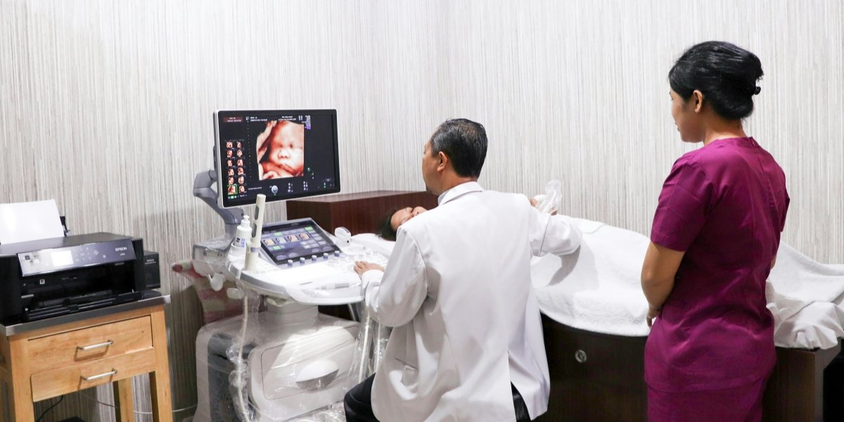
What is an Ultrasound Scanner?
An ultrasound scanner, known medically as an ultrasonic diagnostic apparatus, is a device that uses sound waves with frequencies above the range of human hearing (typically 2-18 MHz) for non-invasive imaging. Its core value lies not only in generating images but, more importantly, in providing real-time physiological and dynamic information – a unique advantage that static imaging like CT or MRI cannot match. According to the World Federation for Ultrasound in Medicine and Biology (WFUMB), due to its zero radiation, real-time capability, and high cost-effectiveness, ultrasound technology has become the most widely used primary imaging modality globally. It is particularly essential for prenatal examinations, abdominal organ assessment, and dynamic cardiac function analysis.
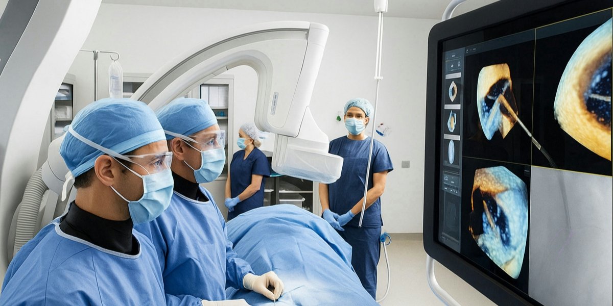
A common misconception is that ultrasound is only for pregnancy checks. In reality, modern medical ultrasound scanners, with their Doppler capabilities, can accurately assess blood flow velocity and direction, making them indispensable for diagnosing vascular diseases and cardiac valve function. The miniaturization of this technology has further spurred the development of handheld ultrasound scanner devices, making point-of-care ultrasound (POCUS) a revolutionary tool in emergency rooms, intensive care units, and general practice.
How Does an Ultrasound Scanner Work?
Its working principle is rooted in classical physics, primarily involving three key steps:
The Piezoelectric Effect and Sound Wave Emission: The core of the probe is a piezoelectric crystal, based on the piezoelectric effect discovered by the Curie brothers in 1880. When an electrical pulse is applied, the crystal vibrates at a high frequency, emitting a beam of ultrasound waves.
Acoustic Impedance and Echo Reception: As sound waves travel through the body, they are partially reflected at interfaces between different tissues (e.g., from soft tissue to bone) due to differences in acoustic impedance. These faint echoes return to the probe and are converted back into electrical signals via the piezoelectric effect. The strength of the echo determines the brightness of the image, while the time of return is used to calculate the depth precisely.
Digital Signal and Image Reconstruction: The breakthrough of modern digital ultrasound scanners lies in the high-speed digitization of analog signals. Through beamforming and parallel processing technology, high-frame-rate two-dimensional or even three-dimensional images are reconstructed in real time. Color Doppler technology goes further by calculating the frequency shift of echoes (the Doppler effect), intuitively encoding blood flow speed and direction with color.
This entire process completely avoids ionizing radiation. Its safety has been confirmed through long-term tracking by the U.S. FDA and the World Health Organization, making it the preferred imaging choice for pregnant women and children.
Who Invented the Ultrasound Scanner?
The birth of medical ultrasound was not an overnight achievement but an epic of collaboration integrating physics, engineering, and medicine:
Theoretical Foundation (18th-19th Century): Italian scientist Lazzaro Spallanzani, in 1794, first systematically explained the principle of echolocation through his study of bat navigation, laying the biophysical groundwork for ultrasound.
Technical Foundation (1880s-1910s): Pierre and Jacques Curie discovered the piezoelectric effect, solving the core problem of sound-to-electricity conversion. Later, in 1915, Paul Langevin developed the first practical ultrasonic transducer for detecting submarines, a technology whose principles are directly linked to medical probes.
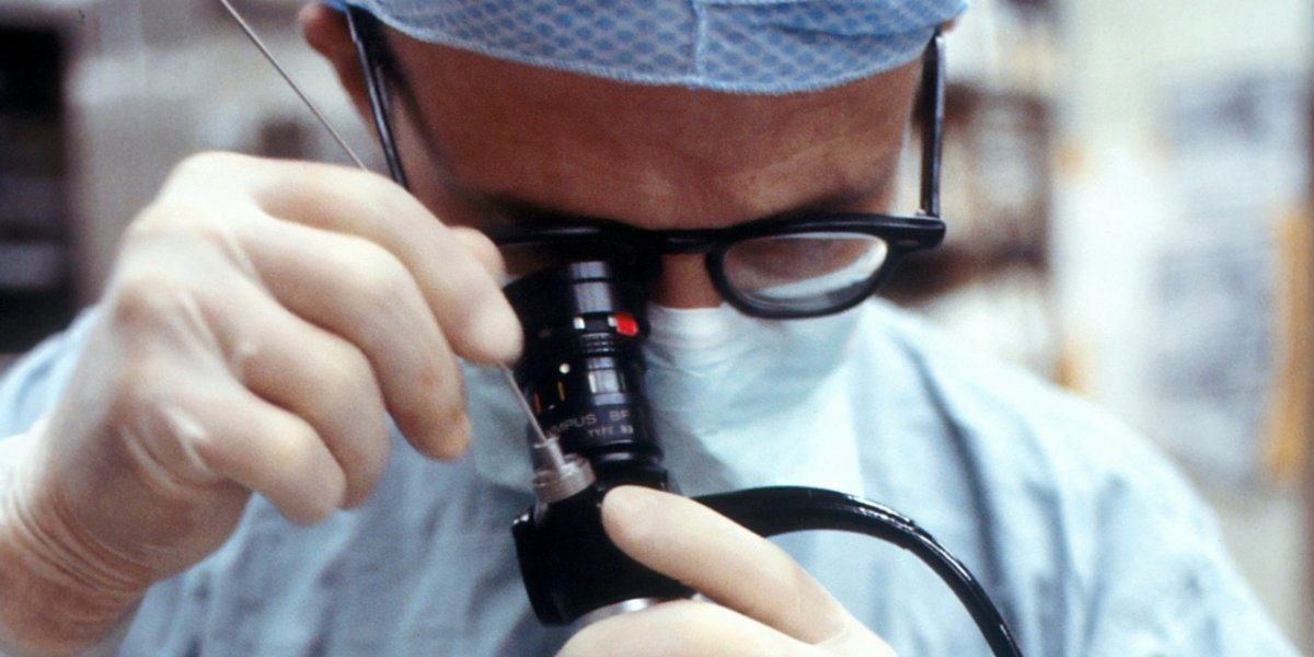
The Dawn of Medical Application (1940s-1950s): After WWII, ultrasound technology shifted from military to civilian use. Austrian neurologist Karl Dussik attempted to use ultrasound to detect brain tumors in 1942, publishing the first medical ultrasound paper—a primitive method but a pioneering effort. The true milestone in clinical translation belongs to the team of Professor Ian Donald at the University of Glasgow, Scotland. In 1958, in collaboration with engineer Tom Brown, they published the landmark paper "Investigation of Abdominal Masses by Pulsed Ultrasound," systematically demonstrating the immense value of ultrasound in diagnosing gynecological tumors, marking the birth of the modern medical ultrasound scanner.
How to Choose an Ultrasound Device for Different Scenarios?
The core principle for selection is "scenario matching," not simply pursuing the highest specifications. Here is an analysis based on clinical needs:
General Practice and Point-of-Care Rapid Diagnosis:
Needs Profile: Rapid screening, high mobility, easy operation.
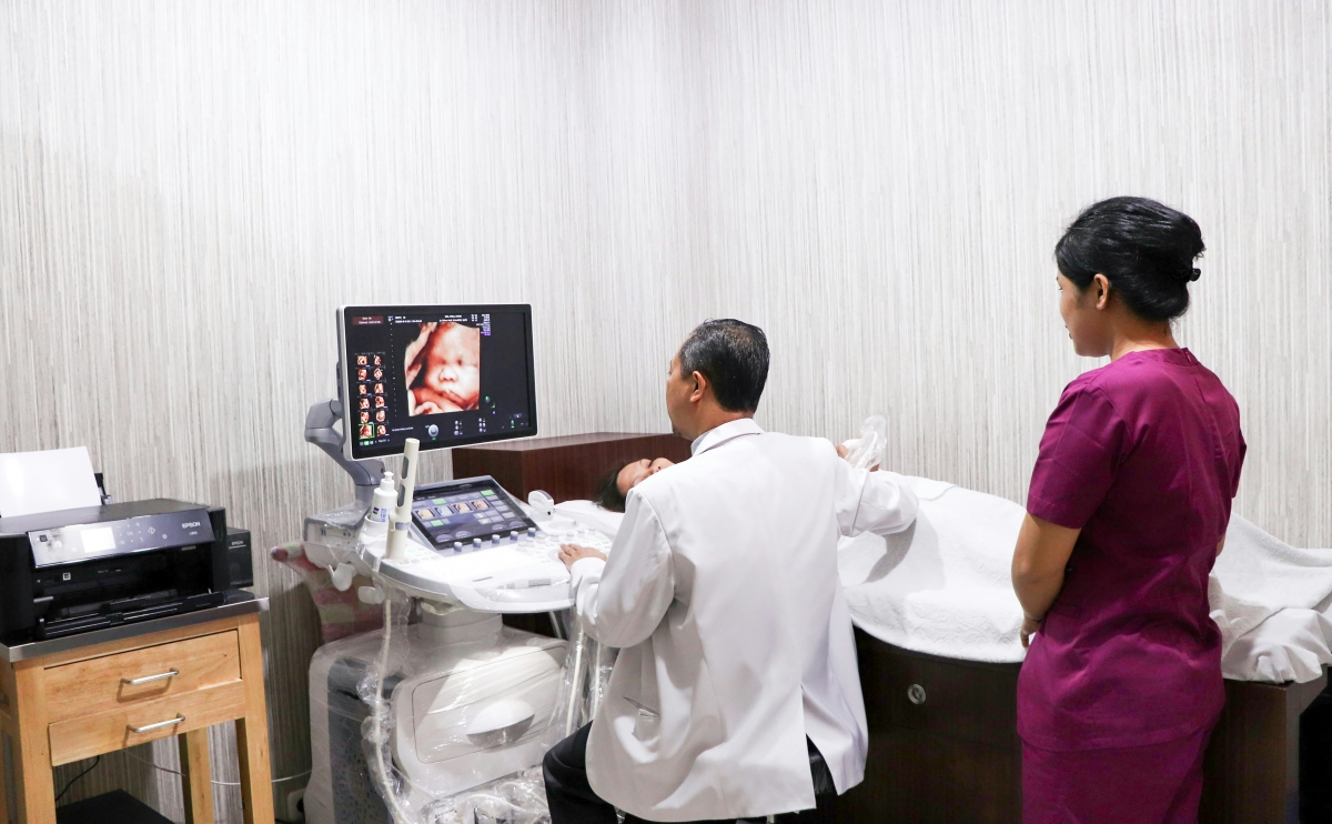
Device Recommendation: A portable handheld ultrasound scanner is the ideal choice. For instance, the Palmsize Digital Ultrasound Diagnostic System WED-2000A. Its design is comparable to a smartphone yet provides clear 2D imaging, making it perfect for home visits, emergency room rapid assessments, and primary care screenings—a true portable ultrasound scanner for pregnancy checks and general use.
Obstetrics/Gynecology Specialty and Vascular Examination:
Needs Profile: High demand for blood flow imaging, requiring fetal anomaly scans, follicle monitoring, or vascular plaque assessment.
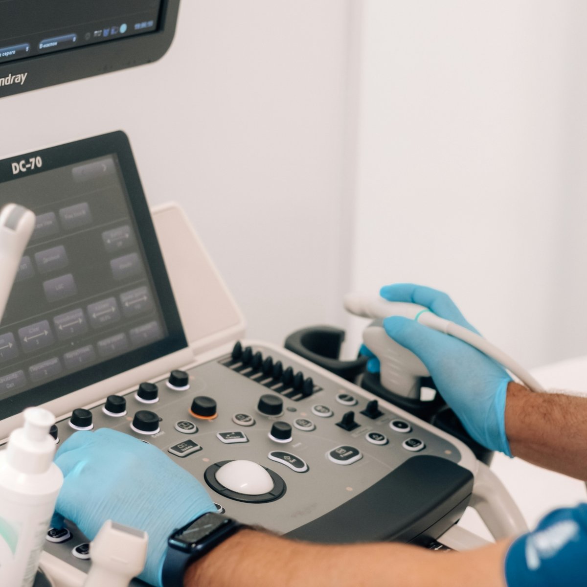
Device Recommendation: Must choose a device with high-performance color Doppler and spectral Doppler capabilities. The Color Doppler Ultrasound System RH-DU8/M9 excels in such applications, providing accurate hemodynamic data to meet the depth requirements of specialty diagnostics—a robust portable color Doppler ultrasound scanner.
Mobile Healthcare and Remote Consultation:
Needs Profile: Highly integrated, lightweight, easy data transfer. Needs to function as a laptop ultrasound scanner or similar.
Device Recommendation: A tablet-based or laptop-based ultrasound system is the optimal solution. The Tablet Specialized Color Doppler Ultrasound WED-WEYE6 combines a powerful processor with a high-definition touchscreen. Images can be shared in real-time via network, making it ideal for telemedicine and rural outreach programs—a versatile mobile ultrasound scanner.
Comprehensive Hospitals and Imaging Centers:
Needs Profile: Shared by multiple departments, handles complex case diagnosis, requires high image quality and comprehensive advanced features.
Device Recommendation: Choose a modular, expandable full digital high-end platform. A system like the All-Digital Ultrasound Diagnostic System WED-180 supports a variety of high-frequency probes for abdomen, cardiac, and superficial organ imaging, equipped with rich quantitative analysis software. It forms the cornerstone for establishing a department's core diagnostic capability—a definitive digital ultrasound scanner workhorse.
Our Expertise
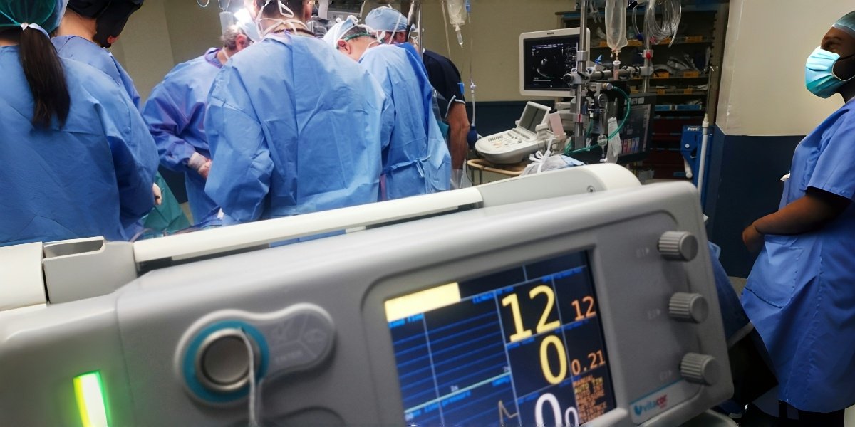
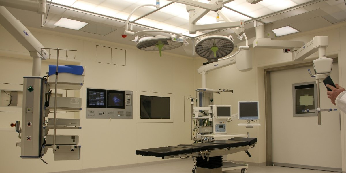
E-Jay Medical is deeply committed to providing reliable, advanced, and efficient integrated solutions for operating theatres and critical care departments. We continuously invest in research and development, keeping pace with the latest trends in medical technology to ensure our products and services meet the increasingly complex demands of modern healthcare environments.
As an innovator in the field of medical technology, we possess profound technical expertise and unique experience in ventilators, anesthesia machines, medical lighting, infusion pumps, and operating theatre solutions. We strive to create a safer, more convenient, and more efficient working environment for healthcare professionals.
E-Jay has become a trusted partner for numerous medical institutions worldwide. Our professional equipment is widely used in operating rooms, ICUs, and emergency resuscitation areas of hospitals at various levels. Supported by a comprehensive service network, we provide customers with timely and professional technical support and after-sales service.